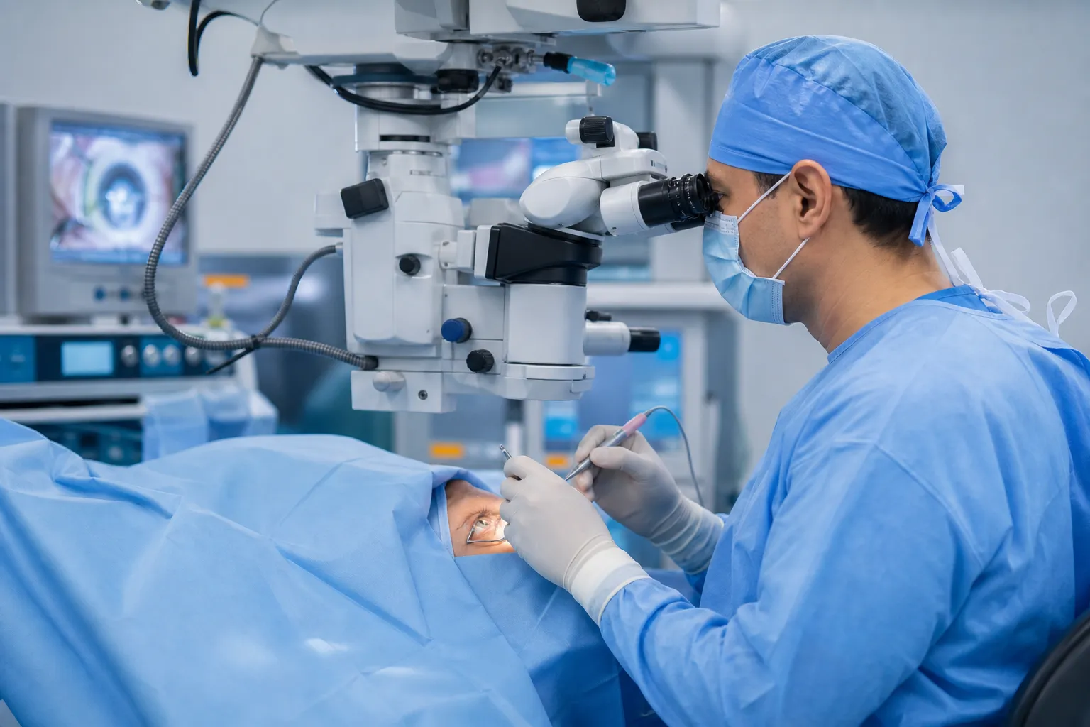
Vitreoretinal surgery
Specialized procedures for retinal detachment, epiretinal membrane, macular hole and complex vitreoretinal pathologies using a microinvasive technique.
What does this service offer?
Ophthalmology Service
Vitreoretinal surgery comprises a wide range of surgical procedures aimed at diagnosing and treating conditions related to the retina and the vitreous, the gel-like substance inside the eye. These surgeries are essential for preserving and restoring vision in patients with various retinal disorders such as retinal detachment, diabetic retinopathy, macular hole, epiretinal membrane and vitreous hemorrhage. Modern small-gauge vitrectomy (23G/25G/27G) allows these pathologies to be treated with minimal trauma to the eye and optimal long-term results.
Why Choose This Service?
Retinal detachment, when the retina separates from its underlying layer, is treated with surgical reattachment to restore its normal function and prevent vision loss.
Diabetic retinopathy, a complication of diabetes that damages the retinal blood vessels, can be surgically repaired, preventing further deterioration.
A macular hole, a break in the center of the macula, is closed by removing the vitreous that pulls on it, restoring sharp central vision.
An epiretinal membrane, a layer of tissue over the surface of the retina that causes visual distortion, is removed with maximum precision.
Vitreoretinal surgery not only preserves existing vision but in many cases restores lost vision, fully reintegrating the patient into daily activities.
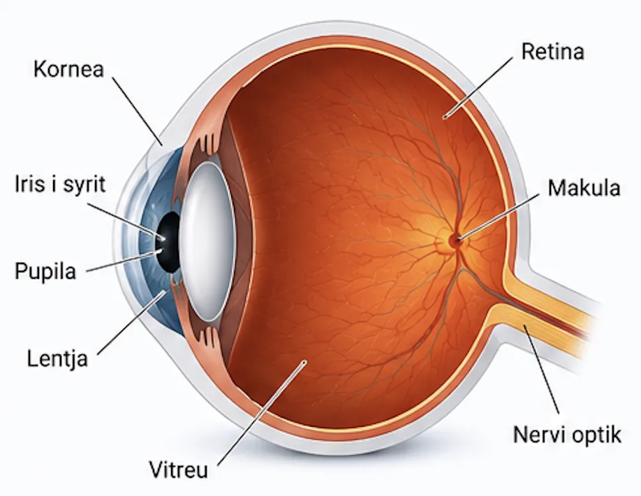
Anatomy of the eye
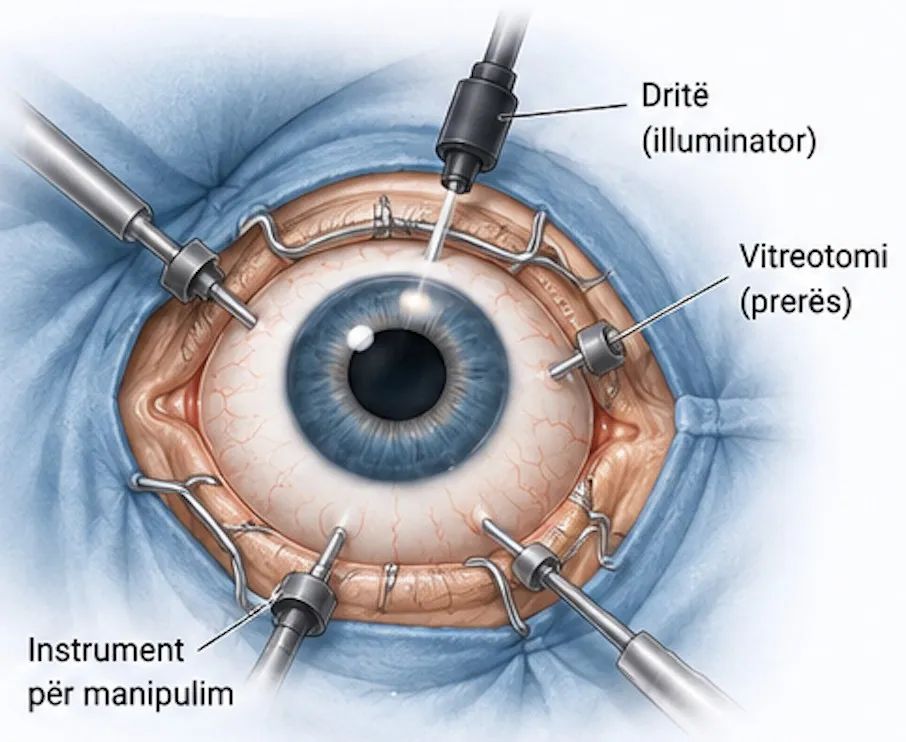
Vitreoretinal surgery instruments
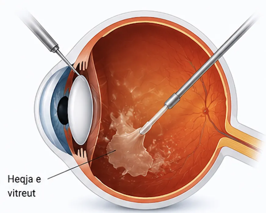
Vitrectomy: removal of the vitreous
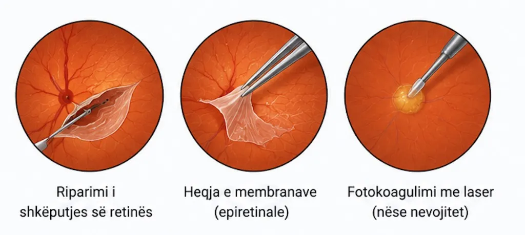
Treatment of the retinal problem
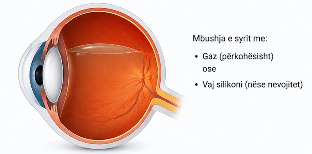
End of the operation: filling the eye
How Is the Treatment Carried Out?
After a detailed evaluation with OCT and ultrasonography, the procedure is performed under local or general anesthesia.
Small-gauge vitrectomy removes the vitreous body, creating direct access to the retina.
Depending on the condition, retinal repair or reattachment, closure of holes and tears, or removal of epiretinal membranes is carried out with high precision.
As needed, a gas bubble (temporarily) or silicone oil is injected to hold the retina in position during healing.
Regular postoperative monitoring ensures full support throughout the recovery process.
Are You Interested in This Service?
Book a consultation with our specialist team to discuss your treatment options.
Book a Visit Now