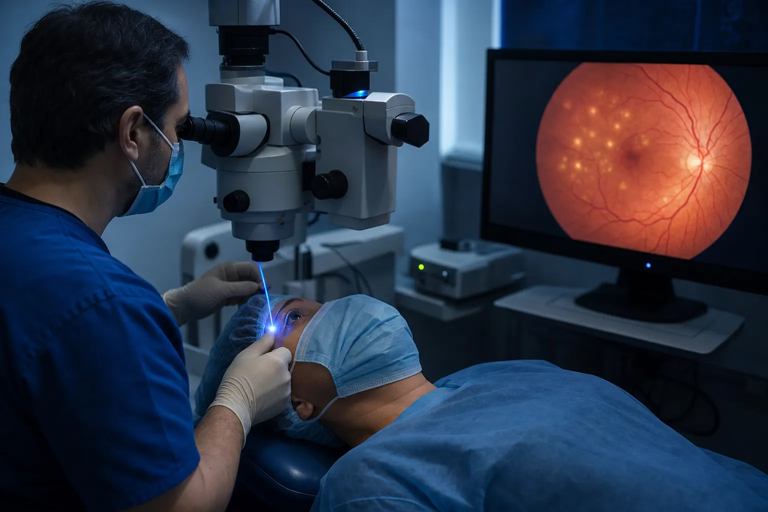
Retinal laser
Laser photocoagulation for the treatment of retinal tears, hemorrhage, edema and diabetic retinopathy — a safe outpatient method with no need for traditional surgery.
What does this service offer?
Ophthalmology Service
The retina is the inner rear layer of the eye and houses the photoreceptor cells that transmit light signals to the brain. It contains the macula, the layer responsible for central and detailed vision, the foveola and the optic nerve. Any damage to this layer, whether from aging, diabetes mellitus, hypertension, trauma or genetic factors, can result in severe visual disorders. Laser surgery harnesses the thermal effect of high-energy beams to treat retinal diseases and is the preferred method when traditional surgery is not necessary.
Why Choose This Service?
Hemorrhages and leakage from retinal blood vessels, secondary to diabetes or hypertension, are sealed and isolated through the thermal effect of the laser.
Retinal edema and macular edema, swelling of the macula that affects central vision, can be reduced by means of laser beams.
Age-related macular degeneration, both its wet and dry forms, is treated by destroying the degenerative areas.
Retinal tears and holes are sealed with thermal energy, forming scar tissue that prevents progression toward complete detachment.
Retinal vascular malformations, abnormal swellings and tortuous contours are also treated with laser.
Laser surgery is far safer than traditional surgery and is performed on an outpatient basis with no need for hospitalization.
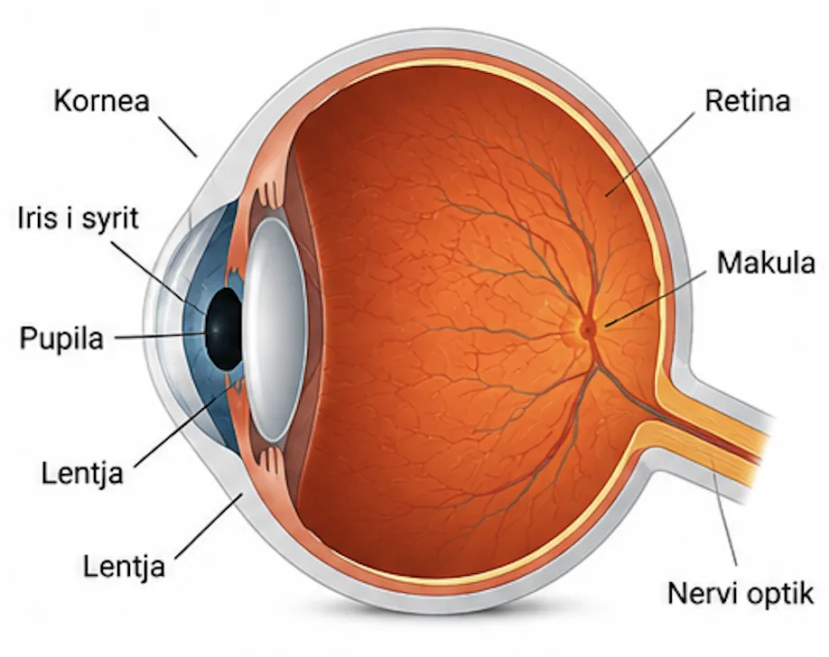
Anatomy of the eye
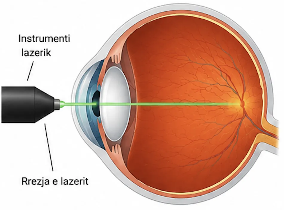
Retinal laser (the principle)
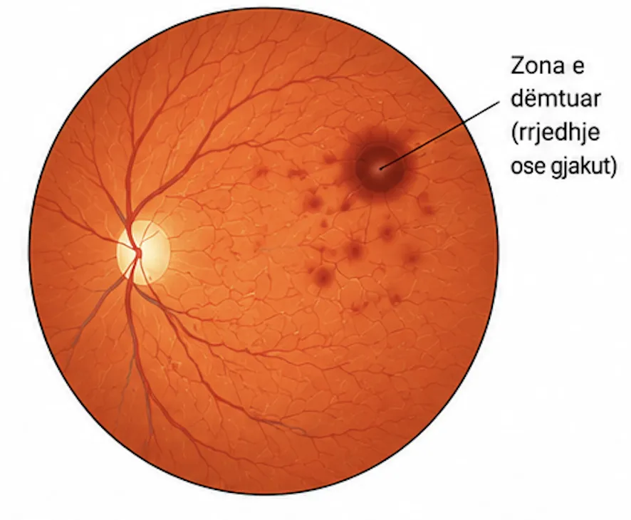
Damaged area in the retina
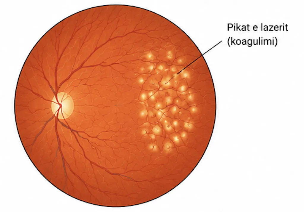
Laser applied to the retina
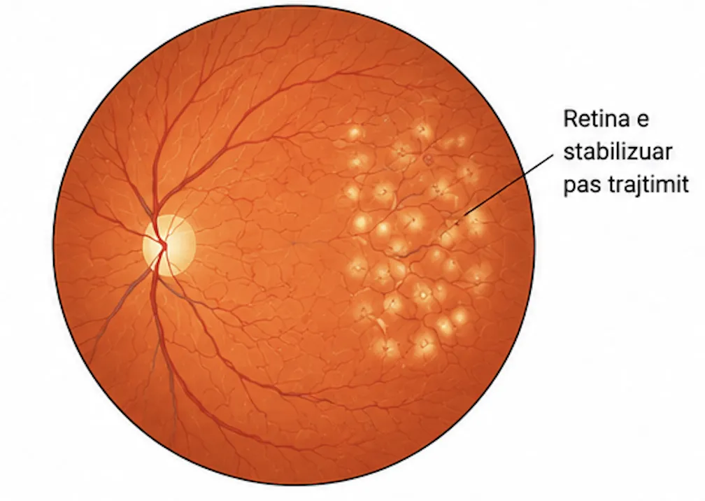
After laser treatment
How Is the Treatment Carried Out?
After a detailed evaluation with fluorescein angiography, optical coherence tomography (OCT) and ocular ultrasonography, the location and severity of the damage are determined.
Before the procedure, stopping anticoagulant medications and starting preventive antibiotics may be required.
The procedure is performed with the patient awake and pain-free: the pupils are dilated with drops and the skin around the eye is numbed.
A special lens is placed on the cornea to focus the laser beams precisely on the diseased area.
The laser beams are fired with control over the hemorrhages, edema, holes or degenerative areas, isolating and sealing the lesion.
The procedure lasts 15–30 minutes; during it, flashes of light may be seen, and afterward mild floaters may be felt, which disappear without treatment.
For one week after the procedure, strenuous physical activity and lifting heavy objects are avoided; the doctor's instructions for antibiotic and anti-inflammatory drops are followed strictly.
Are You Interested in This Service?
Book a consultation with our specialist team to discuss your treatment options.
Book a Visit Now