Pterygium (Surfer's Eye)
A fleshy, triangular growth over the surface of the eye, caused by chronic exposure to UV rays, wind and dust. Treatable with eye drops or surgery.
What does this service offer?
Ophthalmology Service
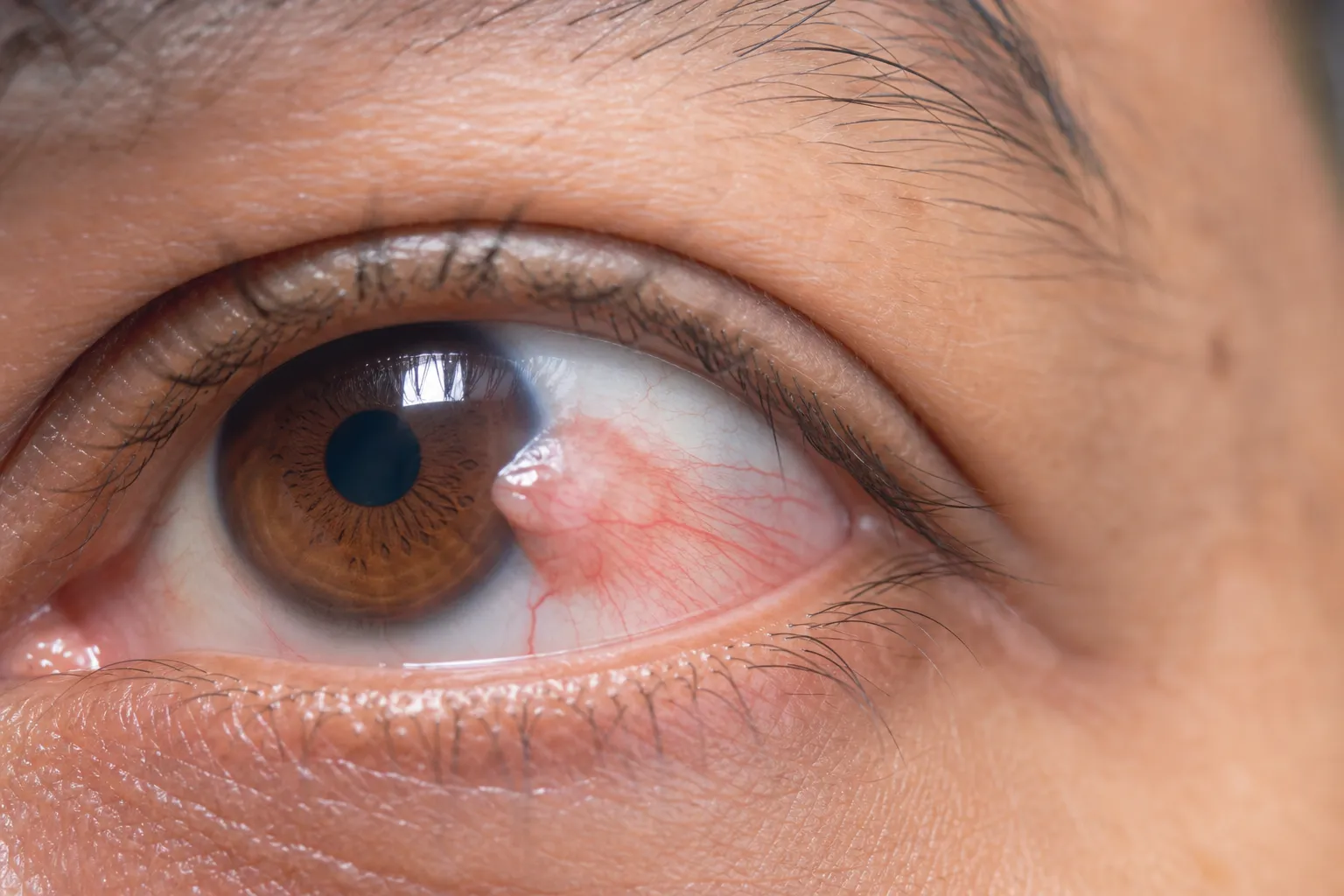
A pterygium is a fleshy, raised growth on the conjunctiva of the eye, the transparent membrane that covers the white part. It may appear white or pink with visible blood vessels and usually begins at the corner of the eye, growing toward the iris. A pterygium is not a cancerous growth and does not spread to neighboring tissues, but it can continue to grow. If it reaches the cornea, it may change its shape, causing astigmatism, or leave scarring that affects vision even after removal. The main cause is long-term exposure to the sun's UV rays, as well as chronic irritation from hot, dry weather, wind and dust. Other risk factors include age over 60, genetic predisposition, vitamin A deficiency and the HPV virus. Symptoms may be completely absent or include redness, a foreign-body sensation, burning, itching, dry or watery eyes and, over time, visual changes.
Why Choose This Service?
Removal of the pterygium prevents further damage to the cornea and restores eye comfort.
Modern surgical techniques using a conjunctival autograft or an amniotic membrane graft offer a low recurrence rate (only 2–15%).
Postoperative treatment with antibiotic and steroid eye drops protects against infection and prevents recurrence.
UV-protective glasses and artificial lubricants can slow the growth of the pterygium before or after the procedure.
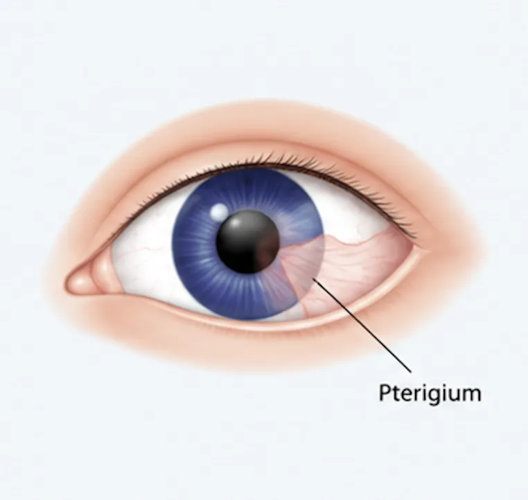
Eye with pterygium
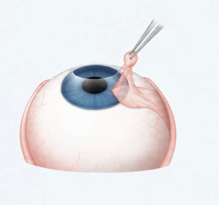
Removal of the pterygium
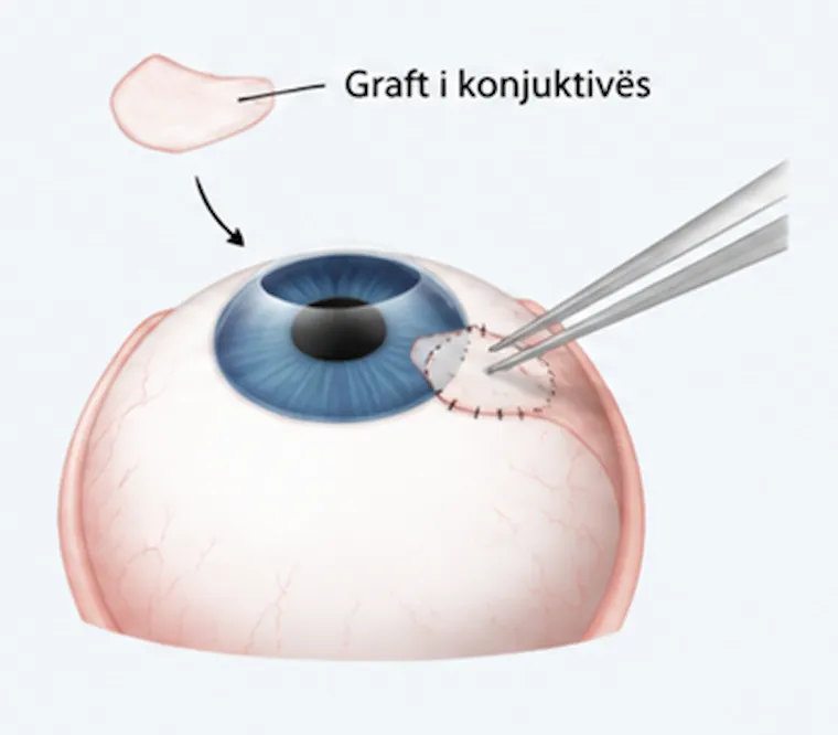
Placement of the conjunctival graft

Eye after treatment
How Is the Treatment Carried Out?
If the pterygium causes no symptoms, the doctor monitors it regularly, usually once a year.
For mild irritation, over-the-counter eye drops or ointment are prescribed, or steroid drops for stronger symptoms.
Surgery is the only way to remove it completely.
During the operation, the ophthalmologist applies local anesthesia, removes the overgrown tissue and covers the gap with an autologous conjunctiva (taken from the patient's own eye) or with donated amniotic tissue, acting as a biological dressing until the conjunctiva regrows on its own.
The operation lasts about 30 minutes.
After the procedure, an occlusive eye patch is applied for 2–3 days and the patient follows a course of antibiotic and steroid drops.
Full healing takes 5 days.
If the pterygium recurs, usually within 4–12 months, a second procedure is performed with additional preventive measures.
Are You Interested in This Service?
Book a consultation with our specialist team to discuss your treatment options.
Book a Visit Now